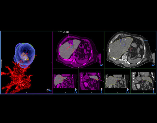
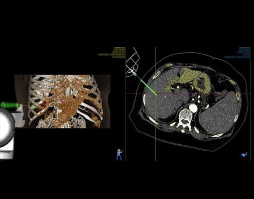
Liver tumor - MWA
MWA 25 mm mass at 8th segment of the liver.
Challenge : Tumor is located close to heart and it is deep seated. MAXIO® helps to plan the safe trajectory path and showing the ablation volume covers the tumor. In the Pre and Post ablation registration – 3D shows the tumor (Orange Colour) is completely covered by the ablated region (Blue colour) as per the planned ablation
Procedure : Needle placement was good and the tumor ablated in single sitting as per plan and in 3D pre and Post ablation registration helps to verify the procedure immediately and the tumor was completed ablated.
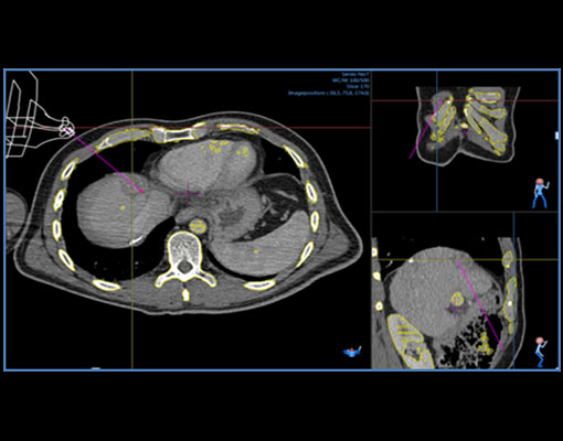
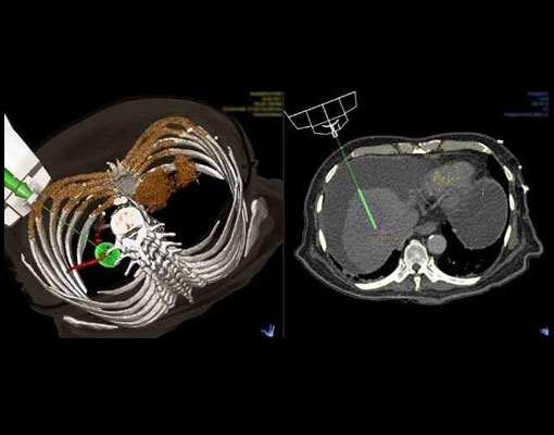
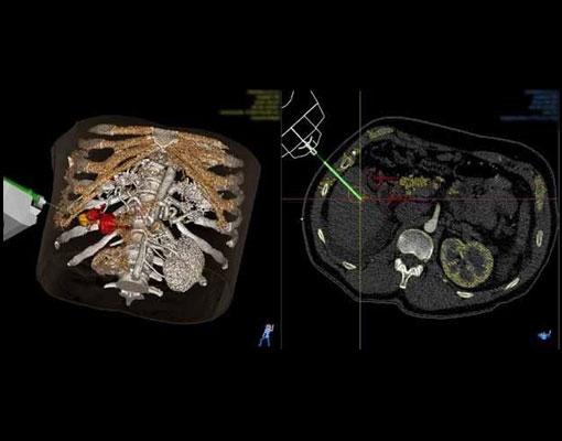
Liver tumor - MWA
MWA 13 mm mass at 5th segment of the liver.
Challenge : Tumor is located close to major portal vein and it is deep seated
Procedure : MAXIO® helps to place the needle precisely and in the Pre and Post ablation registration – 3D shows the tumor (Orange Colour) is completely covered by the ablated region (Blue colour).
Radio Frequency Ablation of recurrent HCC after TACE
A patient was planned for radiofrequency ablation of 3.8 x 3.7 cm lesion at segment 8 of liver
Challenge : Post TACE lesion was not visible on ultrasound. The residual lesion was situated below diaphragm
Procedure : Patient was planned under GA, coolitip: 17G 150/30 Two probes were used.
Orbital angle : 64/63°
CC angle : 18/13°
Depth : 90.9 / 91 mm
Procedure time : 28 min
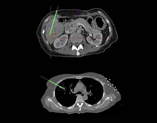
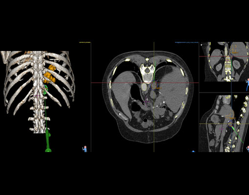
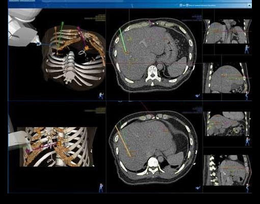
Radio Frequency Ablation of a HCC and a Lung Lesion
Patient planned for RFA of 25 mm mass at 6th segment of the liver and 9 mm mass in right lung during the same procedure
Challenge : Planning safe trajectories with minimal patient discomfort and less procedure related complications is a challenge
Procedure : Ablation procedure planned in two sittings. Lesions targeted precisely with the assistance of MAXIO in sequential mode. No post procedure complications.
Liver plan:
Orbital angle : 64/63°
CC angle : 18/13°
Depth : 90.9 / 91 mm
Procedure time : 28 min
Lung plan:
Orbital angle : -61.84
CC angle : 0°
Depth : - 74.42 mm
Needle used : 17G
Cooltip : 150-30
Planning time : 10 min
Procedure time : 40 m
Radio Frequency Ablation of Adrenal Tumor
Patient diagnosed with HCC underwent liver transplant now with solitary adrenal metastatic lesion
Challenge : In free hand, it is difficult to place the probe with steep angulations to reach the target without traumatising the diaphragm and renal vessels.
Procedure : MAXIO® helps in precise planning and targeting of the adrenal lesion with such complex oblique approach, so as to avoid passing through pleural recess.
Size : 15 mm
Orbital : -5.49°
Cranio caudal : 48.55°
Target depth : 48.55 mm
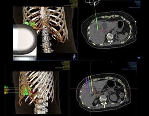
Radio Frequency Ablation of Liver lesion with multiple probes
A patient planned for Radio-frequency ablation with multi-probe placement
Challenge : Multiple probe placement required to cover the lesion completely. Overlapping the ablation zones is a challenge to avoid the residual tumors.
Procedure : MAXIO® helps in multi-probe placement and precise targeting of the lesion. Ablation planned in 5 sittings through simultaneous and sequential mode.
Orbital angle : 7.85°
Depth : 93.83 mm
Needle used : 17G
Cooltip : 150-40
Planning time : 15 min
Procedure time : 80 min
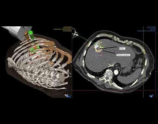
Radio Frequency Ablation of 2 cm of HCC
Patient planned for radiofrequency ablation at 7th segment of liver
Challenge : Lesion is situated posteriorly so planning and treatment is done in prone position.
Procedure : 2 cm lesion was targeted under GA in prone position. The thermal simulation area covered the lesion.
Orbital angle : 19.46°
Depth : 76.28 mm
Needle used : 17G
Cooltip : 150-30
Number of needles : one
Planning time : 10 min
Procedure time : 30 min
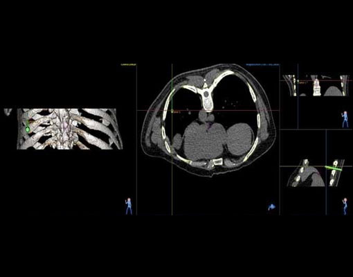
Radio Frequency Ablation of Multiple Lung Lesions
A patient is planned for Radio-frequency ablation of 3 sub-cm lung lesions of primary HCC
Challenge : Precise targeting of the 6mm peripheral lung nodule is difficult.
Procedure : MAXIO® enables better planning for small lung lesions, minimising number of passes / adjustments required for optimal probe positioning. Procedure completed without any complication
Cranio caudal : 9.37°
Target angle : 65.56 mm
Radio Frequency Ablation of 12 mm Mass in Liver
RFA of 12 mm mass at 5th segment of the liver followed by alcohol ablation
Challenge : Ablation area cannot be predicted easily due to its position in the liver segment.
Procedure : MAXIO® helps to visualize the ablation zone, so it can be adjusted to treat the tumor precisely. The site of lesion is well within the ablation zone in post RFA CT.
Orbital angle : 45.6°
Depth : 64.5 mm
Needle used : 17G
Cooltip : 100-30
Number of needles : one
Planning time : 5 min
Procedure time : 22 min
Radio Frequency Ablation 1.8 cm HCC
A patient is planned for radiofrequency ablation of 5th segment of 1.8 cm lesion
Challenge : A deep seated lesion requiring the short and safe trajectory.
Procedure : 1.8 cm lesion A deep seated lesion requiring the is targeted with cooltip probe.
Orbital angle : 23.17
CC angle : 0.97°
Depth : 118 mm
Needle used : 17G
Cooltip : 150-30
Number of needles : 1
Planning time : 9 min
Procedure time : 30 min
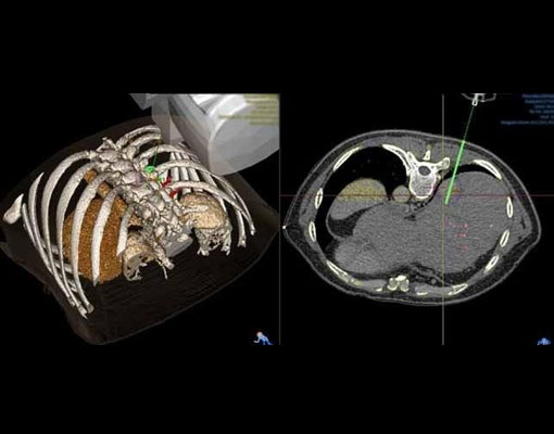
Radio Frequency Ablation of 3.4 cm HCC
A patient is planned for radio frequency ablation of 3.4 cm lesion in 5 th segment of liver
Challenge : Completely covering the tumour with safety margin by thermal ablation zone in sequential probe placements.
Procedure : The lesion is ablated in two sitting of ablation. Two needles placed in simultaneous and first sitting of ablation done and third needle placed towards caudal border by manipulation and second sitting of ablation done.
Orbital angle : 47.08
CC angle : 0°
Depth : 140 mm
Needle used : 17G
Cooltip : 200-30
Number of needles : 2
Planning time : 9 min
Procedure time : 45 min
Radio Frequency Ablation of 1.5 cm HCC
A patient planned for a radiofrequency ablation of 1.5 cm lesion in 3rd segment of liver
Challenge : Avoiding the No Go region like stomach in planning and targeting is a challenge.
Procedure : RFA done under GA.
Cooltip : 150/30
Orbital angle : 9.56°
CC angle : 6.88°
Depth : 100.23 mm
Planning time : 5 min
Procedure time : 15 min
Radio Frequency Ablation of Segment 5 HCC
Patient planned for RFA of (HCC) at Segment 5, with liver cirrhosis and fluid around liver
Challenge : It is a tough case because the lesion, to be targeted, is bouncing due to cirrhosis and fluid around liver.
Procedure : 2 cms HCC targeted for RFA with MAXIO at segment 5 of the liver and RFA performed. Trauma to the bowel was avoided by adjusting thermal simulation option. Post ablation scan shows, a wedge-shaped area without enhancement covering lesion.
Lesion size : 1.6 x 2 x1.3 cms
Orbital angle : 44°
Depth : 69 mm
Needle used : 17 G/100 mm,
Cooltip : 100-30
Number of needles : 1
Planning time : 10 min
Procedure time : 25 min
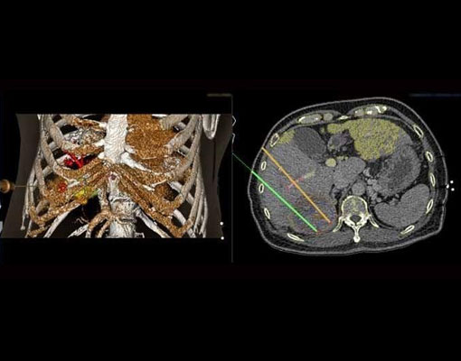
Multiple Liver tumours planned for MWA
- Multiple probe planning in sequential mode
- Overlapping ablation zone can be estimated
- Collision detection and sequencing algorithm

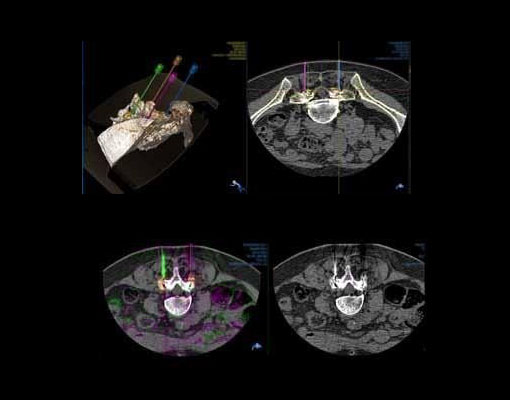
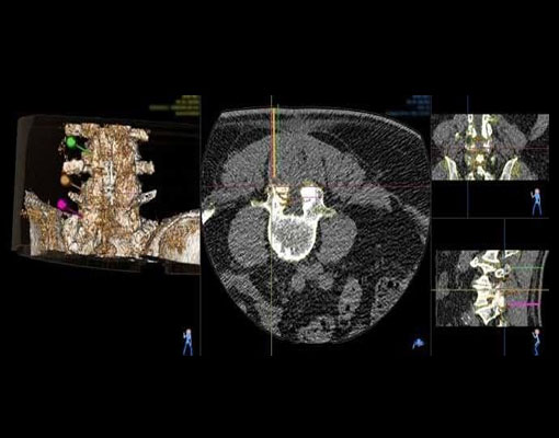
Multiple Facet Joint Injections at L4-L5 & L5-S1
Patient with degenerative lumbar spine planned for facet joint injections at L4-L5 and L5-S1 on right and left side simultaneously
Challenge : Multiple Facet joints at L4-L5 and L5-S1 on right side and left side to be targeted.
Procedure : Facet joint targeted at multiple levels with multiple needles simultaneously, with assistance of MAXIO®. Each Facet joint injected with 1ml of Marcaine and 1ml of Celestone Chronodose. Procedure completed precisely.
Orbital angle : 10°
Depth : 68 to 75 mm
Needle used : 19G/127.5 mm
Number of needles : 4
Planning time : 15 min
Procedure time : 35 min
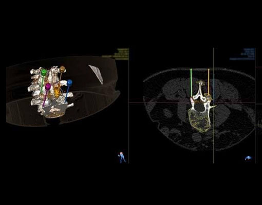
Multiple Facet Joint Injections at L4-L5-S1
Patient with degenerative lower back pain and Non-operative management, planned for facet joint injection at L4-L5-S1 level
Challenge : Multiple Facet joints at L4-L5-S1 to be targeted.
Procedure : Facet joint targeted at multiple levels with multiple needles simultaneously, with assistance of MAXIO®. Each Facet joint injected with 1ml of Marcaine and 1ml of Celestone Chronodose. Procedure completed precisely.
Orbital angle : 0°
Depth : 44 to 52 mm
Needle used : 22G/100 mm
Number of needles : 4
Planning time : 6 min
Procedure time : 31 min for four needles
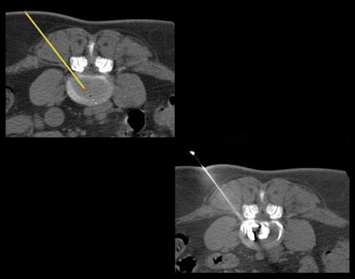
Ozone Injection Into Inter Vertebral Disc
Disc bulge causing compression on L5 nerve root on left side
Challenge : The depth is more and need precise plan to reach the central nucleus of affected disc.
Procedure : Ozone nucleus Injection into the Disc to suck out the water content, after the injection the size of the disc will get reduced in diameter and relieves pressure on nerve roots. The L4-L5 central nucleus of the disc is targeted
Orbital angle : 38°
Depth : 102 mm
Needle used : 18G / 150 mm
Number of needles : one
Planning time : 5 min
Procedure time : 10 min
Facet Joint Injections
Patient with post lumbar decompression and left Para spinal pain planned for multilevel facet joint injections on left side
Challenge : Multiple Facet joints to be precisely targeted.
Procedure : Facet joints on the left side at L3-L4, L4-L5, L5, S1 precisely targeted simultaneously with MAXIO®. Each Facet joint injected with 1ml of Marcaine and 1ml of Celestone Chronodose.
Orbital angle : 0 to 1°
Depth : 58 to 59 mm
Needle used : 22G/100 mm
Number of needles : 3
Planning time : 7 min
Procedure time : 25 min for three needles
Facet Joint Injections
Patient with lower back pain, planned for facet Joint injection at L4-L5 and L5-S1 level
Challenge : Multiple Facet joints at L4-L5 and L5-S1 to be targeted.
Procedure : MAXIO® assisted in precise and simutaneous targeting of facet joint at Multiple level. Each Facet joint injected with 1ml of Marcaine and 1ml of Celestone Chronodose Patient tolerated the procedure well.
Orbital angle : 0 to 1°
Depth : 52 to 60 mm
Needle used : 22G/90 mm
Number of needles : 4
Planning time : 6 min
Procedure time : 60 min